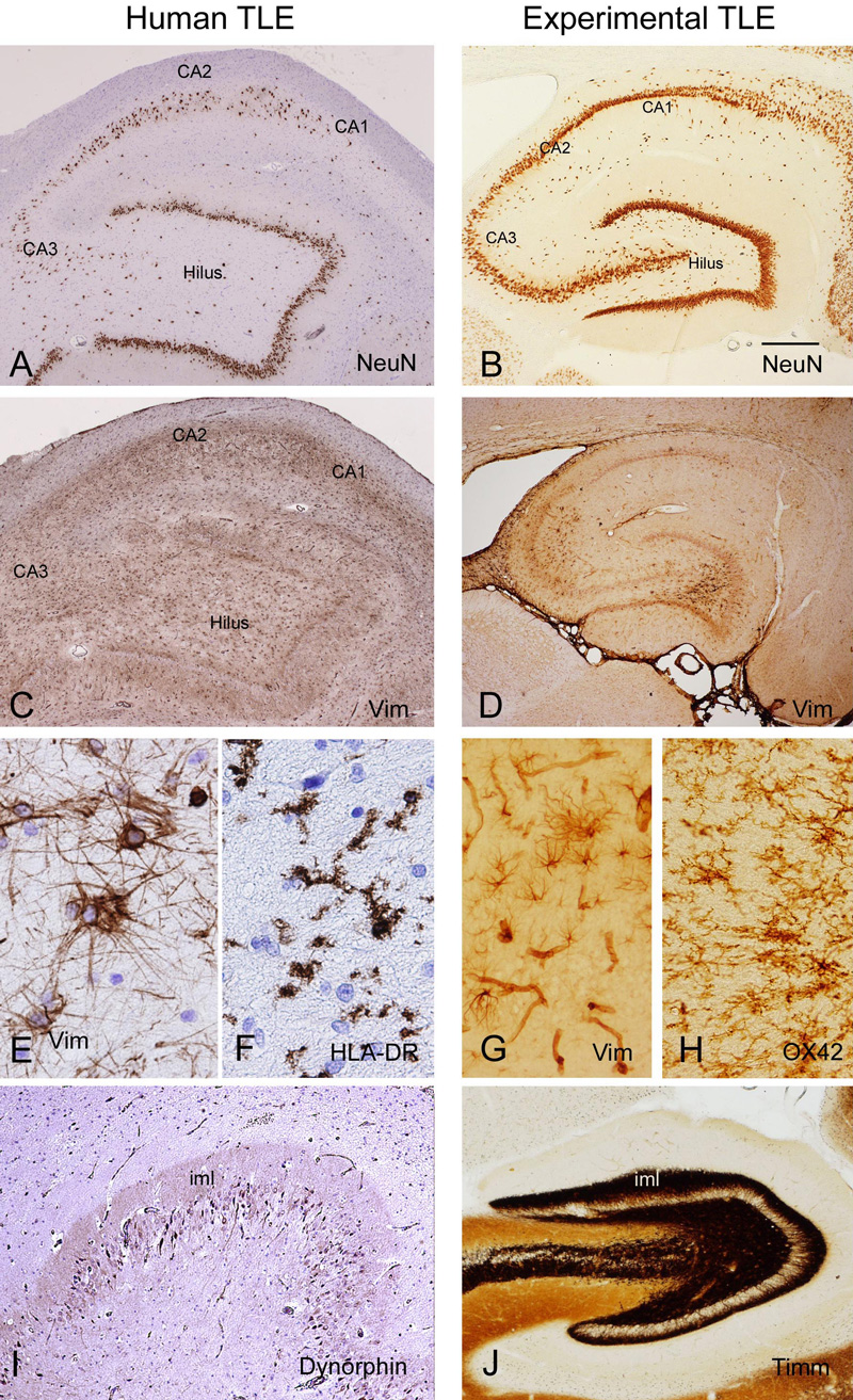
Neuronal cell loss. A and B: neuronal nuclear protein (NeuN) staining showing the loss of pyramidal neurons in CA regions (CA1 and CA3) and in the hilus. Neuronal loss in experimental model (electrical stimulation) is more extensive in hilus than in CA regions.
Gliosis. C, D and E: Vimentin (Vim) staining showing prominent astrogliosis in the regions where neuronal cell loss occurs. F: human leukocyte antigen (HLA)-DR staining showing reactive migroglia cells in the hilar region. F: OX42 staining showing reactive microglia cells in experimental temporal lobe epilepsy. G: Vimentin staining shows reactive astrocytes (and blood vessels) in chronic epileptic rat CA3. H: Ox-42 staining shows microglial cells in chronic epileptic rat CA3 region.
Sprouting fibers. I: Dynorphin staining shows positivity in the molecular layer with a pattern similar to Timm’s staining in experimental temporal lobe epilepsy (J). Bar in B is 300 µm in A and C, 200 µm in B, 380 µm in D, 50µm in E and F, 8 µm in G and H, 250 µm in I and 120 µm in J. (Aronica and Gorter, Neuroscientist, 13(2):100-8)